|
A 78 year old man with history of Type 2 diabetes mellitus and hypertension presents to the emergency department with Right shoulder pain. The paramedics are concerned as they have found a Left bundle branch block on his ECG. On arrival he appears to be in pain but he has a normal complexion and is not diaphoretic. His vital signs are: HR 98, BP 145/80, RR 18, Sats 97% on air, temp 37.2 On further questioning his GP has been treating him conservatively for a pain in his right shoulder for the last month following its sudden development after “jolting” it whilst cleaning the car. He has been otherwise well but the pain in his shoulder has been getting progressively worse and is now debilitating. It is worse with movement but he hasn’t noticed any change with exertion per se. Examination reveals a clear chest, dual heart sounds with no murmurs and a soft non tender abdomen. Examination of the right shoulder reveals a fullness at the sterno-clavicular joint which is painful to palpation, this has been present since cleaning the car. There is no bony tenderness elsewhere. He has full range of movement of the shoulder but all range of motion is painful. His right limb is neuro-vascularly intact. As part of the work up an ECG is undertaken which confirms a LBBB, serial troponin tests are negative and the Chest X-ray can be seen below: 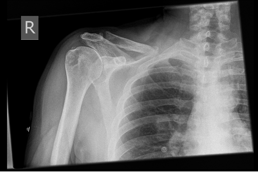 With a keen eye you may notice the calcific deposit in a rotator cuff tendon above the greater tuberosity of the humerus concerning for calcific tendonitis, but did you notice anything abnormal about the clavicle? On closer inspection the proximal half of the clavicle is missing. A CT scan was conducted which demonstrated a soft tissue density mass overlying the medial aspect of the right clavicle which had been eroded with extension into the antero-superior mediastinum. The cause of this mass was a right lower lobe pulmonary primary tumour. This initial Xray finding was sadly missed by the treating doctor and the reporting radiologist, and only when the patient returned due to increasing analgesia requirements a week later was it spotted. Although diagnosis at the first attendance wouldn't have changed the clinical course, admission for symptom control would have been beneficial; the patient required large doses of intravenous opiates to control his pain. This case highlights many challenges in patient diagnostic strategies in todays busy emergency departments. Firstly there had been an emphasis on ruling out myocardial ischaemia as a cause of chest pain, and although this is clearly an important consideration it is also important to keep a broad differential when assessing patients in the emergency department. The harmful effects of cognitive bias and heuristics are important in emergency medicine especially when we are making quick decisions in a busy environment. Be aware of anchoring bias which occurs when the clinician prematurely settles on a diagnosis due to important initial features of the presentation and failing to adjust the diagnostic workup when new information is obtained. This can lead to diagnostic momentum bias which leads the clinician down a diagnostic path, failing to acknowledge other potential causes. In this case the potential for myocardial ischaemia as a cause of the chest pain was given a lot of weight due to the paramedics concern for ACS and the LBBB on ECG. There was also an element of premature closure in this case as once the serial troponins were negative it was thought safe to send the patient home with analgesia and safety netting without consideration for whether there was another rare, life threatening cause. When reviewing X-rays it is important to look at the whole image each time. When its busy we naturally rely on pattern recognition and probability of abnormality from the history and examination. Make sure you have a systematic approach to reviewing all X-rays to ensure that you don't miss rare and not so obvious findings. This patient had a primary diagnosis of lung cancer presenting as a metastatic clavicular mass which is clearly very rare, but it is our job in the emergency department to be able to identify the “needle in the haystack”. Through keeping a broad differential, being aware of our potential cognitive biases and throughly assessing patients, blood tests and imaging methodically we will be less likely to miss these rare but important diagnoses and provide excellent clinical care. For more information on Cognitive bias and heuristics please read: http://www.stemlynsblog.org/its-a-trap/ https://first10em.com/cognitive-errors/ Martin Dore
1 Comment
28/7/2022 16:59:24
https://www.kriptoseyir.com/category/bitcoin-nasil-alinir/ Leave a Reply. |
|
|