|
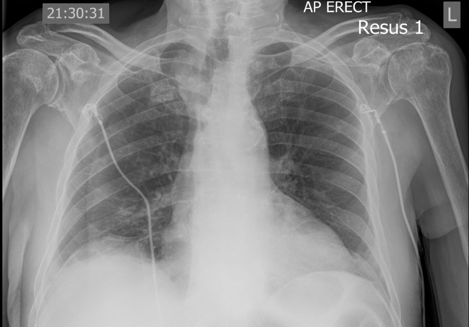
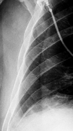
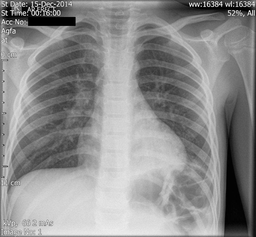
This is the CXR if an elderly gentleman, 83 years, who presented in resus after a collapse. He was found to be SOB and had a CXR: What do you think: does he need a drain? On further inspection and manipulating the x-ray:
6 Comments
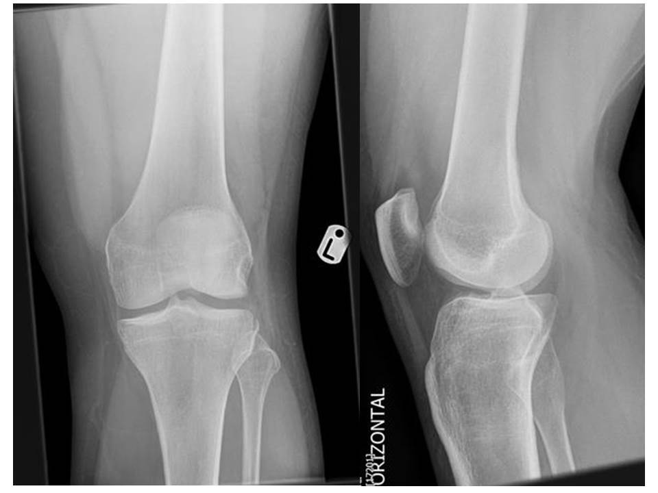
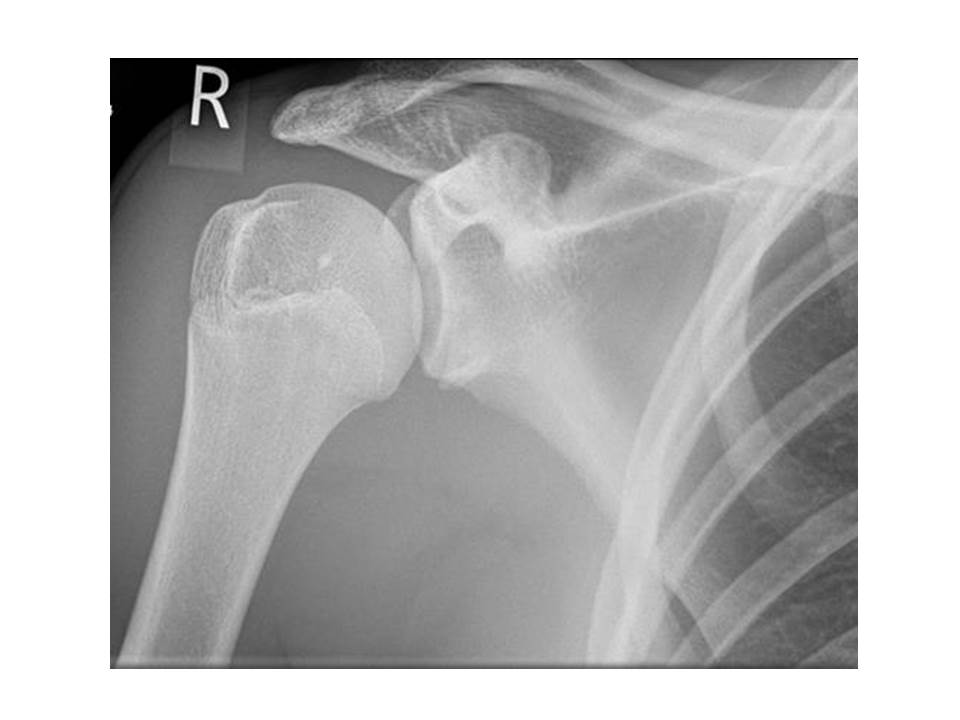
I attended the ED xray meeting last week, and there were a number of interesting cases discussed with relatively subtle xray findings. Remember, anyone is able to attend at 0830 on a Tuesday morning in the Xray seminar room. Patient 1: Missed fracture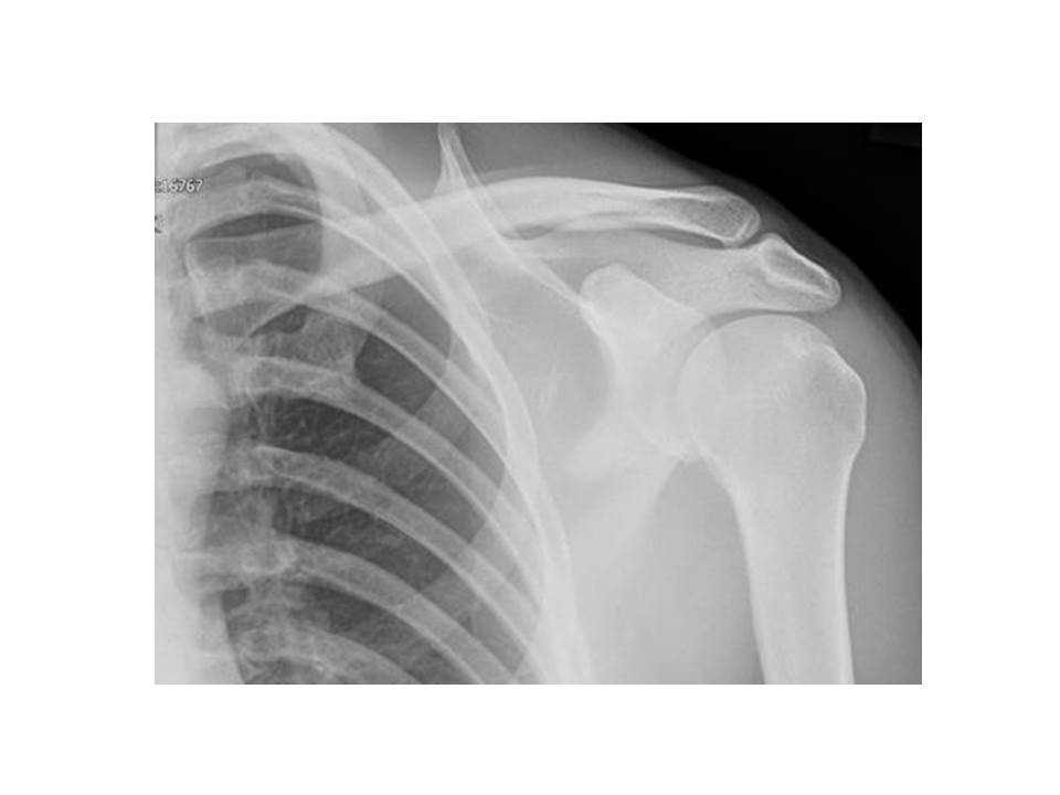 This patient presented to ED following a fall with shoulder pain. She was discharged following XR. The radiologists spotted a fracture and the patient was contacted and re-attended for a CT scan and treatment. Can you spot the abnormality? Patient 2: Segond fracture,This patient presented 2 days after his injury: he experienced sudden severe pain in his knee while dancing. The knee was swollen and he was partially weight bearing. The avulsion fracture at the lateral aspect of the proximal tibia, seen best on the AP view, is a Segond fracture. This type of fracture is often associated with an ACL injury and requires follow up in acute knee clinic. To read more, have a look at the Radiopaedia article here. Patient 3: Bankart lesionNote the fracture fragment projected over the inferior glenoid. This patient sustained a dislocation with resultant bankart lesion. A 'soft' bankart lesion involves only the glenoid labrum, while the less common 'hard' bankart lesion involves a bony fragment. To read more, have a look at the Radiopaedia article here. Clare Bosanko
by Adam Herbstritt6 year old presents unwell, febrile, viral, coryzal with some focal chest signs.
This Xray was performed in ED - what do you see? |
Categories
All
The Derrifoam BlogWelcome to the Derrifoam blog - interesting pictures, numbers, pitfalls and learning points from the last few weeks. Qualityish CPD made quick and easy..... Archives
October 2022
|
