|
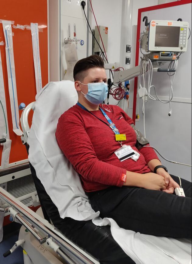
For October the sim theme is “breathing”. This blog covers some of the learning points from 29/10/20. We will be aiming to run simulations weekly - mostly Fridays but not always - see the gmail calendar. November will be "cardiovascular" month. The simulated case: Sam is a woman in her seventies presenting with increasing shortness of breath over the last 2 days. She is requiring >10L/min of oxygen by facemask to keep saturations >94%. At this point consider how wide the potential causes of breathlessness are. After treating the hypoxia, which tests or investigations might increase or decrease the likelihood of it being any of these potential diagnoses? What happened? We ran this short simulation with a nurse, HCA and trainee ACP in the resus area of ED. A history was taken, observations recorded and appropriate oxygen delivered. A range of causes were considered and appropriate investigations (bloods, ECG, chest x-ray) carried out. This simulated patient ultimately would have been found to have bilateral pulmonary emboli (they had increased risk due to metastatic cancer); however, in this short sim the intention was for the patient to be assessed, have emergency management and the right tests thought about. What did we think? In debrief we discussed: History taking: When asking rapid questions to narrow down the differential diagnosis, there is a risk with asking questions in the negative (e.g. “you don’t have chest pain at all?”) that the patient may passively reply “no” to questions, compared to “are you in any pain?” they may be more likely to explain that they do. Investigating and treating PE: We discussed the PERC score, Well’s score, ECG signs, and the treatment options for PE including anticoagulation, thrombolysis and interventional radiology - see guidelines section below. In terms of ECG signs, the most reliable is sinus tachycardia, however this article and its links cover well the signs of right heart strain to look for, and how to differentiate it from similar presentations. Decision making in ED: Breathlessness (or even hypoxia) has a wide list of potential causes. In the emergency department patients are being seen often at the early stages of illness where the disease is potentially less manifest and information is scarce. At this point there is a much higher uncertainty. Prof Carley has a recorded talk and a blog about making decisions amongst this uncertainty here. In cases I have seen during this period of high uncertainty it may be that the patient is treated for several potential causes of their symptoms. For example the patient with PE and secondary heart strain may have already had antibiotics for ?sepsis and dual antiplatelets for ?ACS. This can be okay, as long as the decisions were made with good intentions based on the information available at the time. In the case of those treatments, potentially the benefit from early treatment and the high risk of not treating them may outweigh the risk of giving treatment to someone who is later found not to have the disease. However there are other risks lying in this period of uncertainty. We discussed in the debrief the potential for anchoring bias, where the clinician “anchors” to one early piece of information and all subsequent information is either thought to fit that mental model or is discarded. This may mean that the patient with PE is actually only ever treated for pneumonia, and PE is never considered. Personally, I suspect this bias has greater power when a clinical handover happens - if you are handed over a patient “we’re treating them for X, and they’ve been referred to MAU” is there a risk you anchor to that diagnosis? If new information comes along (e.g. new blood results, their chest x-ray, a colleague reporting they don’t seem to be improving) it’s important to go back and reassess with an open mind. Anecdotally someone I know who suffered a PE explained to me their experience in ED felt like minds had been made up immediately and subsequent information didn’t seem to adjust that idea. Another similar bias we discussed was confirmation bias: believing the patient has a particular diagnosis and then unconsciously only retaining information that supports this, discounting that which refutes it. A technique to combat these biases is to actively seek out information which would change your diagnosis or plan. What other diagnoses would be really important not to miss, and what signs might lead to that diagnosis instead? In our new layout of ED, with front-door senior assessments, patients often have a potential diagnostic label attached to them before they reach the more junior clinicians. This has clear logical benefits for patients. But we raised in debrief that there is the potential for the biases above to occur following this. More junior members of the team may feel difficulty in broaching alternative diagnoses. So we discussed in general how one might explore decision making with a colleague by framing it as a “teaching moment”. For example, “I wouldn’t have thought about X diagnosis for this person, do you mind helping me understand why it is X and not potentially Y?” or “I noticed X piece of information, from my lectures they used to say X was associated with Y, but here you’ve said it’s most likely Z - do you mind telling me about why it’s different here?”. We’ve talked before in this blog about graded assertiveness and the PACE model here.
For a gateway into the larger field of ‘thinking about how we think’ in emergency medicine, go to this blog by Dr Natalie May. And for a deep-dive, I recommend this ebook on how we make decisions in the ED. The guidelines: The EDIS guideline can be found under “adult medicine”, with other helpful resources being the British Thoracic Society guideline (note from 2003) and this LifeInTheFastLane article. These three have been used in the following sections. The PERC rule is a rule-out scoring system for low risk emergency department patients. A score of zero in a low risk patient means <2% risk of PE, which means the risks of investigating most of these patients further would outweigh benefits averaged over the population. It was not possible to use it in this case as it is only for use when the risk of PE is low (e.g. Wells <1). A Wells score is a very important step in the investigation of potential PE as it helps us determine how likely the diagnosis is as a baseline before any investigation (the pre-test probability). We then seek to use examination and tests to change this probability up or down. A patient’s Wells score helps us decide whether a d-dimer blood test will aid us in the diagnosis or not. Because of the test characteristics of d-dimer, where pre-test probability is low a negative d-dimer can help rule out PE, but where the pre-test probability is high a positive or negative d-dimer will not significantly alter the probability of it being PE. Please do look at our EDIS guideline which has a flowchart on when to use PERC, d-dimer and imaging. The patient in this scenario would have gone on to have a CT pulmonary angiogram. It’s worth noting that patients usually need a green (18 gauge) cannula for this. With the diagnosis confirmed there are different possible treatments. Thrombolysis is generally used when there is ‘massive PE’ i.e. with circulatory compromise, or in PE-associated cardiac arrest where a bolus of 50mg alteplase can be used. The patient in this scenario had normal blood pressure and had significant bleeding risks, so thrombolysis was not being considered initially. Interventional radiology can be used to remove clots. If sub-massive (inc heart strain) or massive PE has been detected, discuss with the ED senior or IR directly whether the patient is suitable. The EDIS guideline gives DOAC dosing or weight-based doses of enoxaparin if anticoagulation is being used. There is a separate guideline on the “outpatient pathway” that shows where someone can be safely discharged with treatment vs when admission is more appropriate. To do: Look at the EDIS PE guideline and the separate link for who can be treated with “the outpatient pathway” [ ] When looking after a patient in the next week try to think specifically about possible diagnostic biases and how you might acknowledge and avoid them based on the above [ ] If you took part in the sim, you can use this blog as a starter to reflect on your own experience of it [ ] James Keitley - ED Sim Fellow --------------- For clinical decisions please refer directly to the guidance. This blog may not be updated. All images copyright- and attribution-free in the public domain.
2 Comments
For October the sim theme is “breathing”. This blog covers some of the learning points from 16/10/20. We will be aiming to run simulations weekly - mostly Fridays but not always - see the gmail calendar. The simulated case: Adam is in his 70s and has presented with shortness of breath, fever and productive cough. He has been brought to the Plym (?COVID) area of the emergency department. What considerations are there in where and how we care for patients like this? What is helpful to prepare before the patient's arrival?
What did we think?
In debrief we discussed: Differences in the environment of Plym theatres to be aware of e.g. how to attach oxygen and how to access help. In particular we noted that the tannoy is different to the one for the rest of the department. To seek help one needs to use the white tannoy on the wall to tannoy to the “green desk” of Plym where they can relay the tannoy to the rest of the department if required. Reflecting on the sim perhaps walkie-talkies to facilitate two-way communications between those in resus and those in the green areas would be helpful, especially if the potential runner might be moving around and completing other tasks. It was noted that often the staffing level does not allow for an additional person to be a runner, so perhaps a walkie-talkie worn by a designated person would aid in making sure someone is available when needed. We discussed the difficulty of requesting a doctor to Plym if there is not someone already present. It is generally done through tannoying for “a doctor”. Perhaps if there was a named person each day that can be tannoyed they would be more likely to respond promptly. In terms of collecting samples like the throat swab or blood bottles, we talked about double bag techniques to pass the samples to the green runner. In this case resus was an amber area as was the nearby corridor so a VBG could have been taken directly to the machine still within amber, however blood tests would have needed ICM stickers applied within the area before they were bagged once, and dropped into a second bag held by someone in the green area. We reviewed the geography of Plym including where to don and doff. The guidelines: The choice of antibiotic in potential community acquired pneumonia can be found on our “RxGuidelines” mobile app. See last week’s blog post for the criteria that determine the need for a patient to go to Plym rather than the main ED. To do: Consider going to Plym and conducting a mental run-through of how you would act with a patient in Plym area if you needed to don PPE/collect samples/call specialties/doff without contaminating clean areas [ ] Have a look at the tannoys on the wall of Plym resus and make sure you know how you would access help from there if you needed it [ ] If you took part in the sim, you can use this blog as a starter to reflect on your own experience of it [ ] James Keitley - ED Sim Fellow --------------- For clinical decisions please refer directly to the guidance. This blog may not be updated. For October the sim theme is “breathing”. This blog covers some of the learning points from 08/10/20. We will be aiming to run simulations weekly - mostly Fridays but not always - see the gmail calendar. We previous ran a simulation a few weeks ago of a paediatric asthma case - see the blog post - and this week have reviewed asthma in an adult case. The simulated case: Laura is in her 20s and has presented short of breath. She has previously been admitted to ICU as a result of her asthma. What key questions are important here to work out the cause of breathlessness?
What did we think? In debrief we discussed: Choice of oxygen delivery: with sats almost normal could apply low flow, or initiate 15L/min non-rebreathe and titrate down with assessment. We talked about identification of whether Laura should be considered a possible COVID19 case, which could have implications for safety of those treating her and geographically where in the department she should be looked after in. The Pubic Health England case definition as of 28/09/20 is “new continuous cough or temperature ≥37.8°C or loss of, or change in, normal sense of smell (anosmia) or taste (ageusia)” (Public Health England 2020) however I will find out exactly which criteria we are working from in the Emergency Department and update this paragraph with that information shortly. [EDIT 14/10/20]: the criteria for moving to Plym ED as of 19/06/20 are: fever PLUS acute-onset respiratory symptoms (persistent cough, hoarseness, nasal congestion/discharge, shortness of breath, sore throat, wheezing, sneezing) OR clinical/radiological pneumonia OR anosmia. The patient in this sim had respiratory symptoms so no fever, so unless pneumonia was clinically expected/radiological found, they were appropriate to be outside of Plym. [end of edit]. We discussed the dose of salbutamol nebuliser. Anecdotally I have been told 5mg produces no additional benefit over 2.5mg with greater risk of side effects, although I admit I haven’t seen the evidence of this. A BestBET specific to COPD reviewed one double-blind RCT and found no difference in outcome between 2.5mg and 5mg (Kusre 2010) - note albuterol is the US name for salbutamol. Please do comment below if you have further experience or information about this choice. We talked through analysis of blood gas results for this patient. In particular the importance of noting pCO2 in an asthma exacerbation. We expect it to be lower than normal range. So if in the normal range on an arterial sample this is a worrying sign of impending exhaustion and failure - escalate these patients urgently. Note on the BTS/SIGN guideline, a normal pCO2 of 4.6–6.0 kPa is considered a sign of life-threatening exacerbation, and a raised pCO2 considered near-fatal. We discussed whether d-dimer would be tested in this scenario. We know that in d-dimer testing it is important to consider both the test characteristics, and the pre-test probability of PE. D-dimer has a good sensitivity for PE, but a specificity of around 41% (Perrier et al 1997), meaning that of people without PE, many will still have a positive test. So we need to consider the patient’s pre-test likelihood of PE, such as with Well’s scoring, to decide how a positive or negative test is going to influence that probability before we test it. In practice it may be that blood is being taken before this assessment has taken place, so if we are already performing coagulation tests on the “blue tube” we can consider after our assessment whether to add-on d-dimer or not. We noted in the scenario several times that communication techniques were used to good effect. In an SBAR handover, a key point of a pause followed by “I am concerned because X” with eye contact grabbed the attention of the listener to vital information. Between colleagues use of “are you happy with doing X while I do Y?” summarised tasks that needed to be done whilst ensuring the other person was trained and able to carry out that task, and allowed them the opportunity to “close the loop” in their response. Feedback from the participants noted that it can be difficult to find peak flow meters, and that it would be helpful to have had greater nursing staffing both in terms of caring for patients like this, but also in being able to attend simulation training. The guidelines: Our EDIS guideline on adult acute asthma is the same as page 17-18 of the BTS/SIGN quick reference guide. This gives an overview or both assessment and treatment. It was used in the scenario to categorise the attack as “acute severe” and not yet in the “life-threatening” or “near-fatal” categories. Choice of steroid: the BTS/SIGN guidelines 2019 (page 102) state: “steroid tablets are as effective as injected steroids, provided they can be swallowed and retained. Prednisolone 40–50 mg daily or parenteral hydrocortisone 400 mg daily (100 mg six hourly) are as effective as higher doses.” BTS/SIGN 2019. To do:
Review the quick overview guideline, which is via “ED browser” on EDIS, or page 17-18 here [ ] Reflect on how scary it would be to be admitted with an asthma attack such as this, and how we might consider helping this anxiety in patients we see [ ] If you need to, consider reading an overview of blood gas interpretation - there are many online, for example this one by Geeky Medics [ ] If you took part in the sim, you can use this blog as a starter to reflect on your own experience of it [ ] James Keitley - ED simulation fellow --------------- For clinical decisions please refer directly to the guidance. This blog may not be updated. We aim to run simulations every Friday at 11am, see the gmail calendar for the up-to-date schedule. The sim homepage is derriforded.com/sim where you can see our monthly theme and you can submit suggestions for what we should cover! In September we have covered (click to see the blog summary):
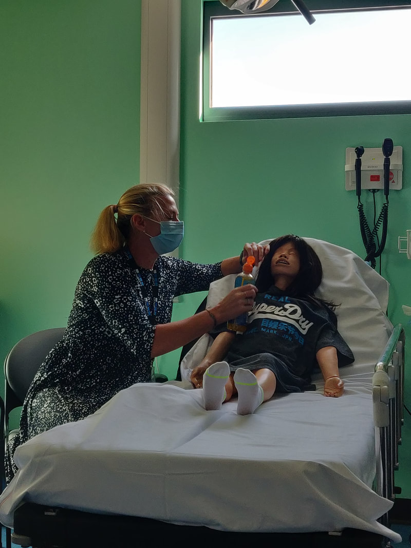
The DKA simulation below was carried out twice and the learning here is a summary of both sessions. The simulated case: Tara is a 10 year old child brought in by a parent. They have been feeling unwell for a few weeks and today they have developed vomiting and pain in their abdomen. Do you already have key diagnoses in mind? What examinations and selective testing will help you rule options in or out?
What did we think? In debrief we discussed:
We discussed in debrief the latest shift in DKA management, from a previous intention to restrict fluid input due to concern of causing cerebral oedema, to a stance of greater fluid administration. The weight of evidence indicates that cerebral oedema develops out of the disease process itself rather than related to fluid-giving. The local guideline (see below) gives clear instruction of the fluid required in this case. We talked through the balance required in keeping the parent informed about what is happening whilst ensuring no delay with immediate care needs. We briefly touched upon the PACE model of assertiveness. This blog describes the challenges of speaking up and how PACE can be used to gradually but assertively escalate your concern to a colleague - you can scroll down to where the example of this is given. Here is a case study of it being used by a “junior” colleague to make suggestions to a “senior” during the resuscitation of a child. Feedback from the sim participants noted the difficulty for adult-trained nurses to be familiar with the paediatric area and equipment - we can aim to pair a paediatric and adult nurse during scenarios. It was suggested we should have more speech directly from the mannequin - we can aim to do this if a facilitator remains outside of the room in future. The guidelines: On the ED browser page you can find two paeds DKA links - the documentation and an appendix for further information. The documentation link is a complete booklet that allows you to write in your results as it guides you through the process and the calculations. You can take a look at the same document on the British Society for Endocrinology and Diabetes page here. It is a good idea to print this off early in the process so it can guide you. I am going to look into whether we can have a few full-colour versions available. This guideline was brought out in March 2020 and represents some significant changes on previous versions that are worth being aware of. As mentioned above, the fluid strategy is now more permissive rather than restrictive, with all patients receiving a fluid bolus of 10mL/kg 0.9% saline, with an extra 10mL/kg (i.e. 20mL/kg total) for those in shock (Tasker 2020). Inadequate fluid resuscitation is noted as one of the key contributors to death in DKA resulting from inadequate cerebral perfusion (BDPED). Have a look at page 5 (and a little at the top of page 7) of the appendix document, also to be found here. This gives a (virtually) single-page overview of what we need to achieve for these patients in the ED. However when you are looking after a patient with DKA you should use the full guideline above. Some more detail on this change in guidance, by Dr Tom Siese: The latest thinking is that rather than the cerebral injury in DKA being simply related to osmotic shifts due to over-rapid fluid treatment, there is increasing evidence which points towards a state of metabolic acidosis and dehydration which then paves the way for a “hyperinflammatory state”. The results from the recent randomised controlled PECARN DKA Fluid Trial (Kuppermann et al 2018) supports the return of permissive fluid boluses in paediatric DKA. At time of writing, the latest editorial in the Archives of Disease in Childhood (Tasker 2020) notes caution in treating cases with altered consciousness, as only 2% of study participants in the above trial had a GCS<14. Therefore the bottom line is don’t be afraid to rehydrate children with DKA, but all cases will still need discussing with a senior member of the paediatric team from an early stage. To do: If it’s been a while since you worked in the paeds area, ask someone who is regularly there to show you where key items are, and if you have supernumerary time you could spend some of it in paediatrics [ ] Have a look at the main guideline document either on our ED browser or via the link above, plus page 5-7 of the appendix document [ ] If you took part in the sim, you can use this blog as a starter to reflect on your own experience of it [ ] James Keitley - ED sim fellow On behalf of the faculty behind this sim Hana Bashir, Andy Robinson, Thomas Siese, Rachel Garlick References:
- British Society for Paediatric Endocrinology and Diabetes [BSPED]. 2020. Integrated care pathway for the management of children and young people with diabetic ketoacidosis. Available from: https://www.bsped.org.uk/media/1742/dka-icp-2020-v1_1.pdf. - British Society for Paediatric Endocrinology and Diabetes. 2020. BSPED Interim Guideline for the Management of Children and Young People under the age of 18 years with Diabetic Ketoacidosis. Available from: https://www.sort.nhs.uk/Media/Guidelines/BSPED-DKA-guideline-2020-update.pdf. - Kuppermann et al. 2018. Clinical Trial of fluid infusion rates for paediatric DKA. NEJM; 378:2275-2287. www.nejm.org/doi/full/10.1056/nejmoa1716816 - Nickson C. 2019. Speaking Up. LifeInTheFastLane blog. Available from: https://litfl.com/speaking-up/. - Tasker R C. 2020. Fluid Management during DKA in children: guidelines, consensus, recommendations and clinical judgement. ADC; 105: 917-918. pubmed.ncbi.nlm.nih.gov/32847796/ - Yianni L, Rodd IG236(P) Pace – ‘Probe, Alert, Challenge, Escalate’ Model of Graded Assertiveness Used in Paediatric ResuscitationArchives of Disease in Childhood 2017;102:A93. --------------- For clinical decisions please refer directly to the guidance. This blog will not be updated. |
Categories
All
The Derrifoam BlogWelcome to the Derrifoam blog - interesting pictures, numbers, pitfalls and learning points from the last few weeks. Qualityish CPD made quick and easy..... Archives
October 2022
|


