|
X-ray request form - "dislocated shoulder and painful jaw." Discharged for follow up of simple dislocation of the shoulder. What are the specific findings on this Xray that show the diagnosis? What else has been missed? 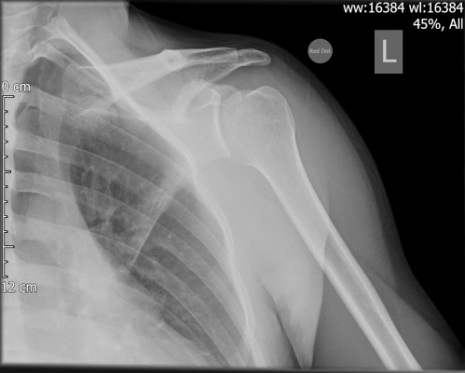 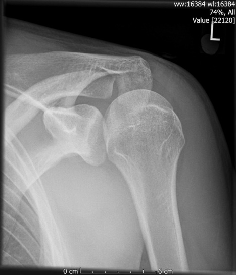 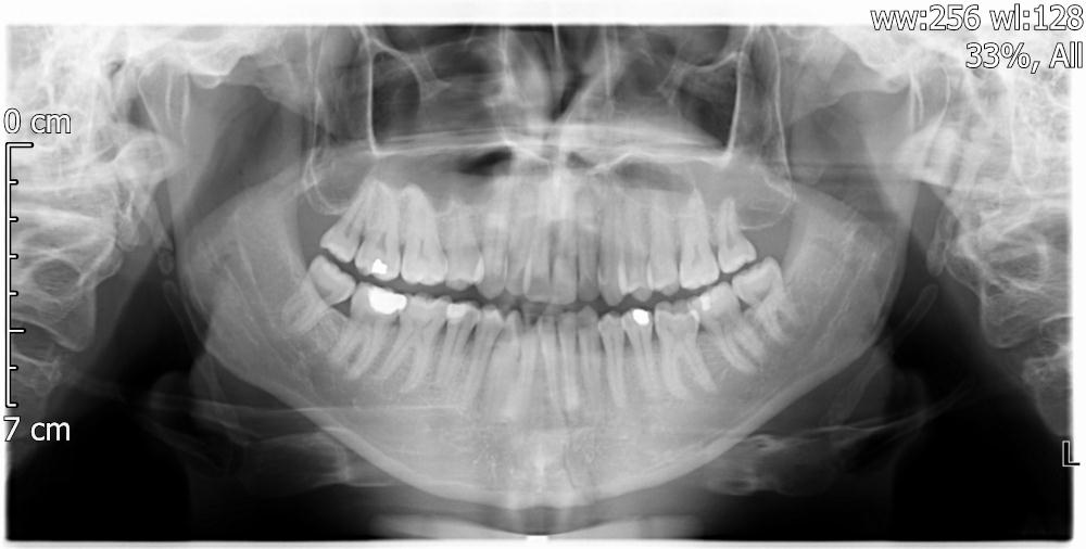 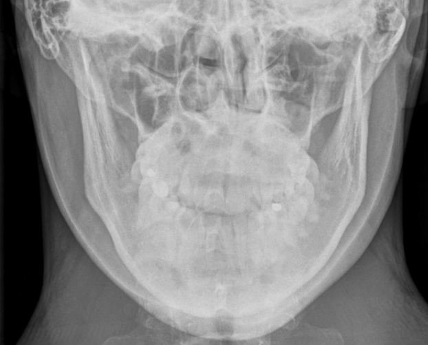 This is a posterior dislocation. They account for perhaps 4% of all shoulder dislocations (apparently commonly associated with fits and electrocution) and are often missed. They usually present adducted and internally rotated. the incidence of neurological complications is lower than for anteriors (as the distance travelled by the humeral head is shorter so there is less traction on the neurovascular structures).
Xrays: Usually there is internal rotation ("Lightbulb sign") and widened glenohumeral space on the AP (loss of the half moon), and loss of contact with the glenoid on the Y view. There is also a suggestion of a "trough line sign" on the AP, which is like a Hill Sachs but on the anterior humeral surface against the posterior glenoid. Classically, reduction of a posterior dislocation is done using a traction technique (which may be in easier in flexion and adduction, with or without posterior pressure on the humeral head). If it is "locked" by the humeral head defect against the glenoid, you can try lateral traction to dis-impact it and then external rotation to unlock it. The missed injury is the jaw fracture on the OPG, just to the left of the midline. 10% of jaw fractures are parasymphyseal like this and about 30% of jaw fractures are unifocal.
0 Comments
Leave a Reply. |
|
|